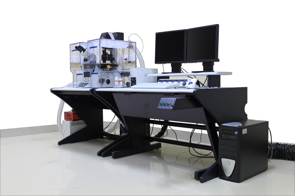
Leica True Confocal Laser Scanning Microscope (CNFL)
- Make
Leica Microsystems, Germany - Model
Leica TCS SP5-II, True Confocal Scanning 5 Channel Spectra Photometer - Specifications
TCS SP5-II for multi discipline use providing high resolution image (8K x 8K) through Motorized Confocal high Speed Scanner. Built in DMI 6000 Inverted Motorized Microscope with stability managerallows us to control intensity of fluorescence with minimum Step size of 15 nm, to get high performance image of Biological Specimens. Various laser sources (He-Ne, Blue diode, light green, red, UV, IR) opticallytunable for confocal optical slicing of fluorescent samples.
Description
Confocal microscopy has become powerful research tool in fields such as Cell Biology, Pathology, Material Science and Nano Sciences etc. it is used in various biological and clinical applications.
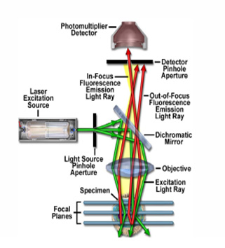
In which Emitted coherent light from laser excitation source passes through a pinhole aperture that is situated in a conjugate plane (Confocal) with scanning point on the specimen and a second pinhole aperture positioned in front of the detector. As the laser reflected by a dichromatic mirror and scanned across the specimen in defined focal plane, secondary florescence emitted from points on the specimen (in the same focal plane) pass back through dichromatic mirror and are focused as Confocal point at the detector pinhole aperture. Collected fluorescence light by the detector translates into electrical signal and multiplied by amplifiers to feed into video display for digitization. Series of images collected from different focal planes of specimen to construct 3-D image Biological/Physical specimen.
Working Principle
Inverted Microscope with built-in stability manager.
Confocal Scanner with large scanning area 22mm to generate highest resolution of 8K x 8K 3-D imaging.
3 Independent Spectral Photo multiplier tubes, each tunable from 400-800nm.
Cooled Photomultiplier and electronics makes system with superior signal to noise ratio and enables system to be used with low laser power on the sample allowing user to keep the cells healthy for long live cell experiments.
Having High power Ar. Laser 100 mw and Ar. Blue Diode Laser 50 mw sources has become extensive requirement to bleach the sample in very short time for bleaching/photo activation experiment because bleaching can be done only by high power laser.
Confocal laser scanning microscope has become powerful research tool in cell biology, clinical studies, Biochemistry, Nanotechnology, Biophysics, digital 3D imaging studies etc.
Biological
Live cell imaging with using fluorescence emission of the sample, which is a powerful contrast technique.
Provides high resolution image with new imaging techniques FRET, FRAP etc.
Study of the cells in Nervous System (Neurobiology).
To study of Micro-organisms and their action on living tissues.
Provides direct visualization of Chromosomes and laser micro dissection allows research in DNA Analysis.
Regular inspection of Growing cells i.e. Cell Culture Studies.
Allows scientific Study in plant science to determine structure, growth, metabolism and deceases of plants.
Clinical examination of cells, tissue specimen and organs allows to determine failures in immune function, it covers immunology.
Bio-physics
To determine molecular scale and structure
To observe multidimensional molecular interaction with help of fluorescence imaging.
Users require submit sample on Poly –l-lysine Microscopic Slide or acid washing cover slips.
User is also allowed to submit the tissue or cell specimen in liquid state.
Before submission of the sample store sample under wet conditions to prevent loss for sample cells.
It gives high performance 3D image of fluorescent Specimen, which belongs to any field of research such as cell biology, cell culture, plant science, microbiology, genetics, immunology, pathology, material science, nanotechnology etc.